Skip to main content
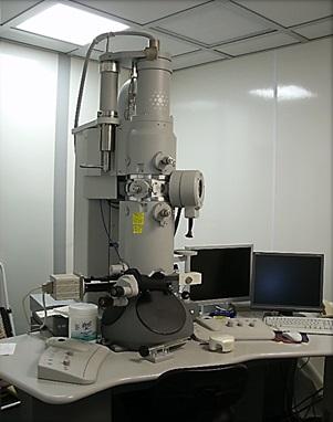
(1) FEI BioTwinG2 Transmission Electron Microscope (Life Sciences building)

- Acceleration voltage of 120kV.
- Goniometer/stage tilt capability.
- Side-mount AMT XR-60 CCD digital camera system for acquiring digital images.
- Film capability
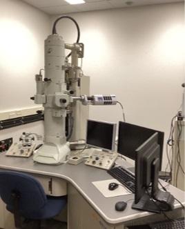
(2) JEOL JEM 1400 Transmission Electron Microscope (AERTC building)

- La B6 filament
- Resolution: 0.2nm/0.38nm(Lattice Image/Point Image)
- Accelerating Voltage up to 120kV
- Magnification(Mag mode/Low Mag mode): x5K-2M/x120-4K
- Specimen Stage: microactive goniometer with piezo drives
- Specimen Chamber(Specimen per Load/specimen Tilt Angle): 1/±25°(±70° with optional holder)
- Equipped with Anti-contamination Device and Cryofin
- Bottom-mount Gatan Orius SC1000B CCD camera
- Equipped with energy dispersive X-ray and Oxford X-MAX 80T detector
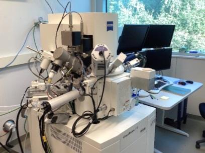
(3) Zeiss Crossbeam 340 focused Ion Beam-Scanning Electron Microscope (FIB-SEM, AERTC building)

- FE-SEM, High Vacuum or Variable pressure mode available
- Multiple detectors available: InLens Duo(SE and BSE), SE2, VPSE
- Capella FIB column with Ga-Liquid metal ion source
- Resolution at 30kV:3nm ,Voltage range:500V-30kV, Probe Current range: 1pA-100nA
- Equipped with Micromanipulator, capable of TEM lamellas specimen preparation
- Gas Injection System of Platinum precursor
- Oxford EDAX and EBSD detectors
- Equipped with Leica Cryo system
- Equipped with Atlas5, a powerful integrated software, capable of 3D tomography imaging and nano-patterning process
(4) Sample Preparation (Life Sciences building and AERTC building)
- Leica EM UC7 Ultramicrotome
- Leica EM UC7/FC7 Ultramicrotome and Cryo-ultramicrotome
- Freeze Plunger FEI Vitrobot
- High Vacuum Coater/Freeze Fracture Unit Leica EM ACE 600:
- Equipped with cryo-transfer SEM sample holder(Leika VCT 100) for Cryo-SEM sample prep
- Turbo Freeze Drier EMS 775
- Cryo Transfer TEM Specimen Holder(Gatan Gat-626)


